
Last updated: 2026-09-11
What Is a Lumbar Disc Herniation?
A lumbar disc herniation occurs when the nucleus pulposus (the soft, jelly-like core inside a spinal disc) tears through the outer annulus fibrosus and pushes out to press on a nerve root. Most cases improve without surgery. Although the problem originates in the lower back, the compressed nerve travels down into the leg, so leg tingling and pain can be more noticeable than back pain itself. (Zhang AS et al., 2023)
A spinal disc consists of a gel-like nucleus pulposus at its center, surrounded by several layers of the annulus fibrosus. The nucleus pulposus helps distribute load, while the annulus fibrosus wraps around and supports it. As people age, the disc loses water content and elasticity, and cracks can form in the annulus fibrosus, allowing the nucleus pulposus to push outward.
Repeatedly bending the lower back, lifting heavy objects, or sitting for long periods puts strain on the discs. Repeated strain layered on top of age-related degenerative changes can trigger symptoms, sometimes starting right after a specific movement.
An analysis of Korean national health insurance claims data found that the number of patients treated for lumbar disc herniation increased until 2012 and then declined, while the proportion of older patients continued to rise. (Jung JM et al., 2020)
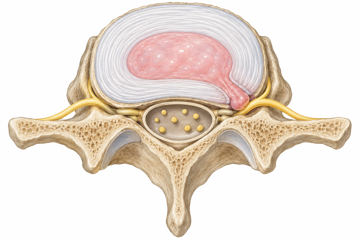
Cross-sectional diagram showing the nucleus pulposus of a disc pushing past the outer annulus fibrosus toward a nearby nerve. AI-generated medical illustration
If Your Back Doesn't Hurt but Your Leg Tingles, Which Nerve Is Compressed?
Tingling that runs from the buttock down to the toes, even when the lower back itself isn't very painful, happens because the location of the symptom differs from the location of the nerve compression. Nerve roots exiting the lower spine control sensation and movement in the legs. When the nucleus pulposus presses on one of these nerve roots and inflammation develops around it, tingling and pain appear in whichever part of the leg that nerve serves. Pain that radiates from the buttock down the leg along a nerve's sensory distribution is called radicular pain, or radiating pain. (Zhang AS et al., 2023)
In common posterolateral herniations, a lesion between the 4th and 5th lumbar vertebrae typically involves the L5 nerve root, which can cause tingling running from the outer calf toward the top of the foot and big toe. A lesion between the 5th lumbar vertebra and the 1st sacral vertebra typically involves the S1 nerve root, with symptoms in the back of the calf, the outer foot, and the sole serving as clues.
Because the direction of herniation determines which nerve is compressed, and because skin sensory zones overlap, tingling on top of the foot narrows down which nerve to examine, but pinpointing the exact nerve requires checking muscle strength and reflexes as well.
Some people feel a sudden jolt of leg pain when coughing or sneezing, or worsening tingling after sitting for a long time. Others feel relief after walking for a short while. Which area becomes uncomfortable after how long of sitting, and how symptoms change with a change in posture, are useful clues for identifying the cause.
How Do You Tell This Apart From Spinal Stenosis, Piriformis Syndrome, or Facet Joint Pain?
The cause of leg tingling can often be distinguished by how far a person can walk, what happens when sitting, and what happens when bending forward or backward. Spinal stenosis commonly causes leg discomfort when standing or walking that eases when sitting or bending forward. Lumbar disc herniation, by contrast, often worsens leg symptoms when bending forward or sitting.
If there is deep pain in the buttock that worsens with sitting, piriformis syndrome is also considered. Tenderness around the buttock and whether symptoms change with hip movement are useful clues, and the straight leg raise test often does not reproduce tingling in these cases.
If pain around the lower back appears when bending backward or twisting, facet joint pain is also evaluated. Facet joint pain can also cause referred pain that spreads to the buttock or thigh. Beyond whether the leg itself hurts, sensory loss following a nerve's distribution or weakness in a specific muscle are what distinguish a disc herniation from these other causes.
New difficulty urinating, urinary incontinence, problems controlling bowel movements, or numbness in the perineal area (the area between the genitals and anus) call for an emergency room visit that same day. Weakness in both legs is another warning sign of cauda equina syndrome. This is a surgical emergency involving severe compression of multiple nerve roots at once, so anyone who suspects it should go to the emergency room immediately; the literature suggests decompression within 24–48 hours. (Kögl N et al., 2024)
Foot drop, in which a person cannot lift the front of the foot and it drags while walking, or rapidly progressing muscle weakness, should also not be delayed. Regardless of how severe the pain is, a new inability to generate muscle strength requires prompt attention to protect nerve function.
What Does THE WELLS Nowon Check?
At THE WELLS Nowon, the physical examination comes first. This includes the straight leg raise test to see if it reproduces the usual leg symptoms, checking which skin areas have reduced sensation, testing strength in the ankle and big toe, and checking knee and ankle reflexes. Based on the exam, the clinician estimates which nerve root's function has declined and distinguishes between cases limited to sensory changes and those that also involve muscle weakness. (Zhang AS et al., 2023)
Next, X-ray imaging shows bone alignment, the space between vertebrae, and bony changes such as bone spurs. While the neurological exam evaluates nerve function, X-ray shows the structure of the bones that make up the lower spine.
The Sigma spinal segmental stiffness test uses a computer to measure resistance at each spinal segment, showing which segment has stiffened and is restricting movement. Musculoskeletal ultrasound shows real-time images of damage and inflammation in surrounding ligaments, tendons, and soft tissue.
If a patient already has an MRI from another hospital, it is brought in and compared against exam findings. Because disc changes are commonly seen on imaging even in adults without symptoms, treatment decisions are based on whether the side with tingling and weakness matches the compressed nerve seen on imaging. If they don't match, other causes or accompanying problems are investigated.
When symptoms stem from nerve root compression, a nerve block can reduce pain in the affected area. If there is also damage to surrounding ligaments or tendons, or muscle tension, prolotherapy injections and manual therapy address those issues as well.
How Does Step-by-Step Treatment Progress, and When Should Surgery Be Discussed?
Many people worry about surgery right away, but conservative treatment comes first unless there is a neurological deficit such as muscle weakness. About 60–80% of patients with lumbar disc herniation improve within 6–12 weeks, and conservative treatment during this period is recommended when nerve function is preserved. (Kögl N et al., 2024)
Korean claims data also show that the number of patients receiving conservative treatment for lumbar disc herniation with leg tingling increased by more than 30% between 2010 and 2019, confirming that a conservative-treatment-first approach is also reflected in clinical practice in Korea. (Cho S et al., 2023)
THE WELLS Nowon addresses acute pain first. Medication is used to control pain, movements that severely aggravate leg symptoms are reduced, and daily movement that pain allows is maintained. If leg pain still interferes with sleep and movement, a nerve block guided by real-time C-arm imaging delivers medication near the compressed nerve root, along with anti-inflammatory IV fluids to calm inflammation and pain.
Anyone taking blood-thinning medication should disclose this before the procedure, since it relates to bleeding risk at the injection site, and some patients notice temporary changes in sensation after the procedure that later resolve.
Once pain decreases, treatment shifts to restoring spinal alignment and movement. Sigma Tapping correction uses a computer to identify stiffened segments and gently taps to correct them, while manual therapy uses hands-on techniques to release stiff segments and tense surrounding muscles, reducing strain when moving the lower back.
If ligament or tendon damage is also confirmed, prolotherapy or ozone injections guided by ultrasound may help support recovery of the damaged tissue, and shockwave therapy is used if tension remains in surrounding muscles or tendons.
Winback/Limpar therapy uses radiofrequency energy to release stiff fascial adhesions and improve range of motion. Because someone with residual leg pain from nerve compression needs different treatment than someone whose stiff surrounding tissue is limiting back movement, the treatments used are matched to the problems identified during the exam.
If pain and functional limitation that interfere with daily life persist despite adequate conservative treatment, and the nerve suspected on exam matches the compression seen on imaging, it's time to discuss surgery. Because nerve recovery declines the longer symptoms persist, cauda equina syndrome, foot drop, and progressive muscle weakness call for discussing surgery right away, without waiting out the full course of conservative treatment.
An observational study following 505 Korean patients with lumbar disc herniation who received integrative Korean medicine treatment for about 52 months reported that 68.4% remained free of symptom recurrence during the follow-up period. (Lee J et al., 2017)
Once pain subsides, the focus shifts to preventing recurrence: breaking up long periods of sitting, lifting objects close to the body, and gradually increasing walking and core strengthening exercises based on how the legs respond.
References
- Zhang AS, Xu A, Ansari K (2023). Lumbar Disc Herniation: Diagnosis and Management. Am J Med. PMID: 37072094
- Jung JM, Lee SU, Hyun SJ (2020). Trends in Incidence and Treatment of Herniated Lumbar Disc in Republic of Korea: A Nationwide Database Study. J Korean Neurosurg Soc. PMID: 31408926
- Kögl N, Petr O, Löscher W (2024). Lumbar Disc Herniation—the Significance of Symptom Duration for the Indication for Surgery. Dtsch Arztebl Int. PMID: 38835174
- Cho S, Lim YC, Kim EJ (2023). Analysis of Conservative Treatment Trends for Lumbar Disc Herniation with Radiculopathy in Korea: A Population-Based Cross-Sectional Study. Healthcare (Basel). PMID: 37628549
- Lee J, Kim J, Shin JS (2017). Long-Term Course to Lumbar Disc Resorption Patients and Predictive Factors Associated with Disc Resorption. Evid Based Complement Alternat Med. PMID: 28769985
Frequently Asked Questions
Q. If leg tingling has improved, can numbness still linger longer?
Yes, numbness can persist even after tingling has improved, since not all symptoms recover at the same pace. If the area of numbness expands or leg strength weakens, a repeat neurological exam is needed even if pain has decreased.
Q. Do I need to stay in bed the whole time with a lumbar disc herniation?
Brief rest is fine when pain is severe, but prolonged bed rest is not recommended. Short walks and changes in position, within a tolerable range, should continue as part of daily activity.
Q. Why do I need to disclose my current medications before a nerve block?
Blood-thinning medications are related to bleeding risk at the injection site, so the name and dosage of any such medication should be disclosed before the procedure. Don't stop any medication on your own — discuss with the prescribing doctor whether and when to pause it.
Q. How should exercise and return-to-work intensity be decided after pain decreases?
Rather than immediately returning to a previous activity level once pain decreases, it's better to start with light activity and gradually increase time and intensity. Consider how much sitting or heavy lifting work involves, and if tingling clearly worsens after activity, reduce activity level and see a doctor to reassess.