
Last updated: 2026-11-05
How Are Spondylolysis and Spondylolisthesis Different?
Spondylolysis is a defect in the bony ring at the back of a vertebra, while spondylolisthesis is a condition in which an upper vertebra has slipped forward relative to the vertebra below it. For patients without emergency neurological symptoms, nonsurgical treatment starts with activity modification and progresses to pain treatment for the surrounding tissue and exercise-based rehabilitation.
Spondylolysis is a condition in which the bony ring at the back of the vertebra, which links the joints above and below, has cracked or broken, leaving a defect. The alignment of the vertebrae above and below, however, stays in place.
Spondylolysis is a stress fracture of the bony ring that develops from repeated backward bending of the low back or forceful twisting of the trunk, rather than from a single major impact. It occurs most often at the fifth lumbar vertebra, which structurally bears the greatest load, followed by the fourth lumbar vertebra. Symptomatic spondylolysis appears more often in people with frequent trunk rotation and extension, such as young athletes. According to a review of the literature, about 80% of symptomatic spondylolysis cases have defects in the bony ring on both sides (Debnath UK, 2021).
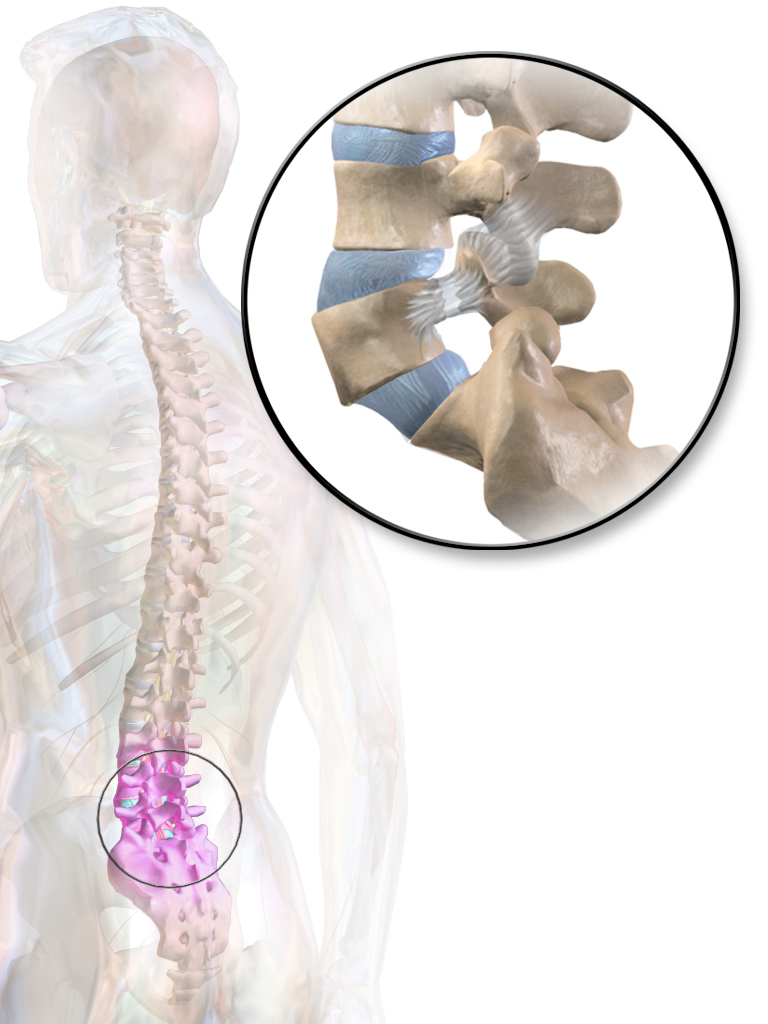 A break in the bony ring at the back of a vertebra. The vertebrae above and below remain in place (BruceBlaus, CC BY-SA 4.0)
Spondylolisthesis refers to a vertebra that has slipped forward, and it is classified as isthmic or degenerative depending on the cause. Isthmic spondylolisthesis occurs when a vertebra loses its support and slips forward because of the bilateral bony ring defects described above. In degenerative spondylolisthesis, by contrast, the bony ring is not broken and remains continuous. However, as the disc (the cartilage between vertebrae) and the facet joints at the back undergo age-related degenerative changes, the spinal segment loosens and the vertebra slips forward (Bydon M et al., 2019).
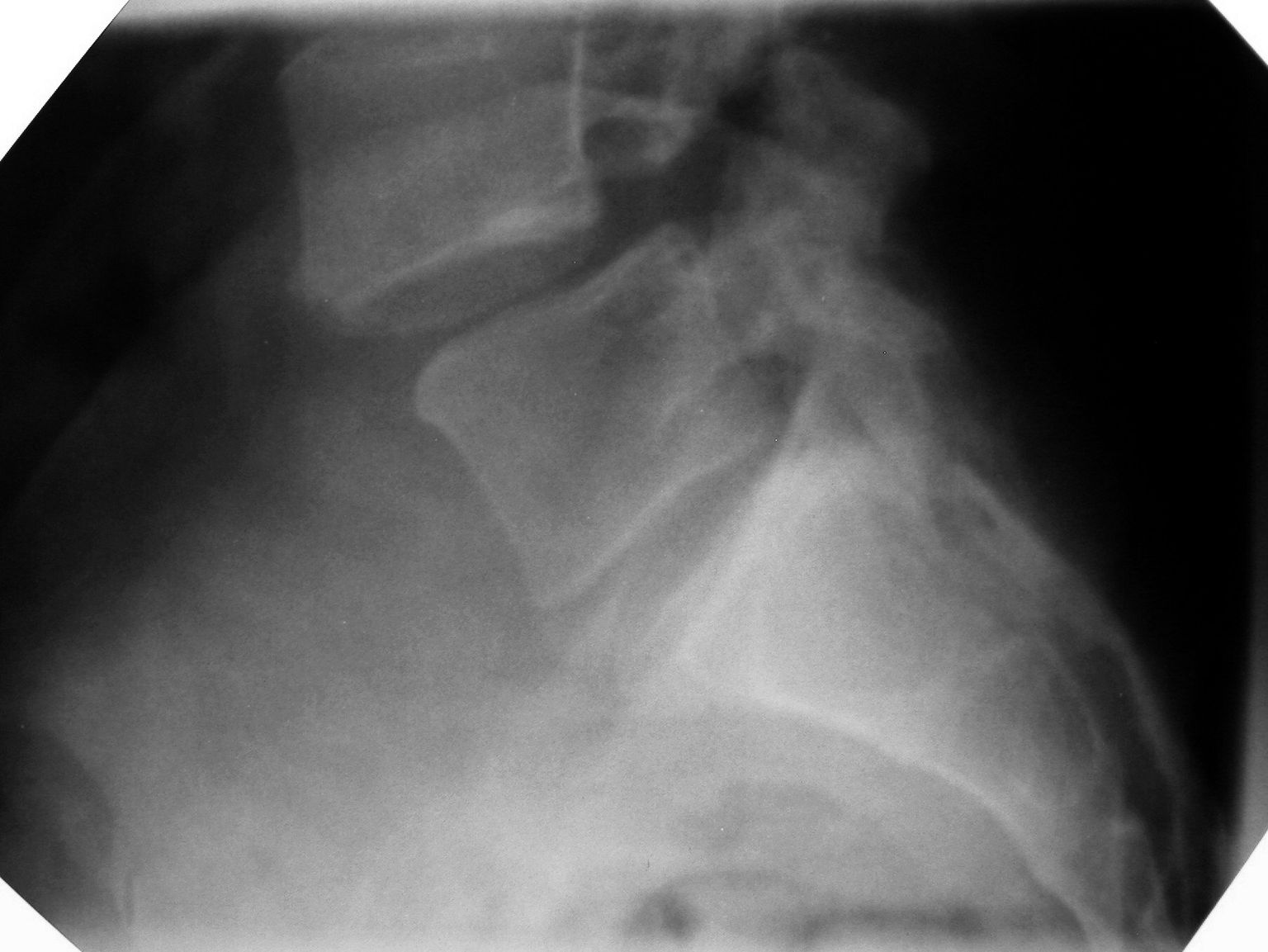 Publicly available lateral lumbar X-ray. The upper vertebra has slipped significantly forward relative to the vertebra below (Jojo, CC BY-SA 3.0)
Symptoms also vary by age and cause. In children and adolescents, spondylolysis feels fine in everyday life, but back pain worsens with exercise or when arching the back backward. The hamstring muscles at the back of the thigh may also become tight and stiff (Cavalier R et al., 2006). Spondylolisthesis, which is common in adults, by contrast, can cause pulling pain in the low back, buttocks, and legs during prolonged standing or walking, but sitting down or bending the low back slightly forward — as when pushing a cart — opens up the space and eases the pain considerably.
Does Spondylolysis Mean the Vertebra Will Keep Slipping?
Long-term follow-up studies show that having a bony ring defect does not mean the vertebra keeps slipping in everyone — in many cases, the vertebra's position stays stable for life.
One long-term study followed 30 first-grade children with a confirmed bony ring defect, out of 500 children screened, for 45 years. Among those with a defect in only one bony ring, not a single person developed vertebral slippage over the 45 years, and their original spinal alignment remained unchanged. Even among those with defects on both sides whose vertebra did slip, the rate of progression clearly slowed with each passing decade, and by the final follow-up, every subject's degree of slippage remained under 40%. The degree of slippage progression also showed no clear association with the low back pain these subjects reported in daily life, and the health questionnaire scores they gave 45 years later were similar to those of the general population in the same age group (Beutler WJ et al., 2003).
Fatigue damage to the bony ring progresses through stages, starting with an early stress reaction and advancing to an incomplete fracture and then a complete fracture. A bony ring defect on only one side tends to have a milder clinical course than a defect on both sides (Debnath UK, 2021).
That said, in children and adolescents going through a growth spurt, spondylolysis and spondylolisthesis require ongoing monitoring — with clinical exams and imaging to check for progression of the slippage — until bone growth is fully complete (Cavalier R et al., 2006). Degenerative spondylolisthesis, commonly seen in adulthood, is itself one of the major causes of low back pain in adults. When the slipped vertebra and a thickened ligament cause stenosis of the spinal canal or of the foramen where nerves exit, this can produce radiating pain with numbness from the buttock down the leg, along with difficulty walking — the legs feel like they're about to burst after walking only a short distance, forcing the person to stop and rest (Bydon M et al., 2019).
Some Spondylolysis Doesn't Show Up on X-rays
THE WELLS first checks the overall alignment of the vertebrae and the joint spacing with an X-ray. An X-ray taken while the patient stands shows how far the upper vertebra has slipped forward relative to the vertebra below it under body weight, while dynamic X-rays taken with the lower back bent forward and arched backward show abnormal movement at that spinal segment. THE WELLS also uses musculoskeletal ultrasound to check for damage and inflammation in the ligaments, tendons, and soft tissue around the lower back in real time in the exam room.
However, plain radiographs alone cannot reliably detect all early-stage bony ring damage. In a clinical study that performed both X-ray and MRI on 200 young athletes with low back pain, MRI detected active spondylolysis in 97 patients—48.5% of all subjects—that the initial X-ray had not revealed (Kobayashi A et al., 2013). This study also found that several physical examination findings investigated together did not show statistically significant predictive power for early detection of spondylolysis.
Based on this evidence, THE WELLS recommends an MRI to check for early bony ring fatigue damage in young athletes whose X-ray is normal but whose low back pain persists. In adult patients with spondylolisthesis, narrowing of the spinal canal and nerve pathways can also cause radiating pain or neurogenic claudication—leg numbness that occurs while walking (Bydon M et al., 2019). Accordingly, THE WELLS recommends MRI to precisely assess the degree of nerve compression in adult spondylolisthesis patients who have leg numbness, difficulty walking, or leg weakness.
My Legs Feel Weak and I Can't Urinate Properly
If you can't urinate even though your bladder is full, if you suddenly lose control over urination or bowel movements, if the skin around your groin and inner buttocks (the saddle area) feels numb as if it belonged to someone else, or if both legs suddenly give out and you collapse, you should go to the emergency room that same day. These symptoms—urinary dysfunction, reduced sensation in the saddle area, and a sudden change in muscle strength—are emergency warning signs that occur when the bundle of nerves at the lower end of the spinal canal is strongly compressed all at once, as seen in cauda equina syndrome or severe nerve compression.
Even if your symptoms aren't severe enough to require an emergency room visit, if your ankle or big toe won't lift properly while walking so that your foot keeps catching, or if one leg has been getting progressively weaker than before, nerve damage may be underway. In that case, you need prompt medical evaluation.
In pediatric and adolescent patients, if pain that interferes with daily life persists even after adequate non-surgical treatment, or if follow-up imaging shows continuing progression of the slippage or of neurological deficits, surgical consultation becomes appropriate (Cavalier R et al., 2006). In particular, when symptomatic children and adolescents have high-grade slippage exceeding 50%, it becomes harder to predict how well they will respond to non-surgical treatment. In adults with degenerative spondylolisthesis, if symptoms such as pain and difficulty walking persist even after consistent conservative (non-surgical) treatment, decompression surgery alone—which widens the compressed nerve pathway—or decompression combined with spinal fusion becomes an option in a surgical consultation (Bydon M et al., 2019).
From Rest to Prolozone: Step-by-Step Nonsurgical Treatment
Without emergency nerve paralysis or severe instability, most patients with spondylolysis or low-grade spondylolisthesis can continue nonsurgical treatment, dividing it into stages with different goals over time. The sequence starts with reducing the mechanical stress placed on the bone, then relieving pain in the overloaded surrounding ligaments and fascia, and finally training the muscles that support the spine on their own.
In the acute phase, when back pain first begins, activities that arch the back backward or strongly twist the torso should be reduced above all else. For early stress reactions in the bony ring in children and adolescents, conservative treatment involves briefly pausing exercise to limit activity and immobilizing the spine. In practice, spondylolysis in children and adolescents often heals well with a short period of activity restriction, immobilization, and physical therapy. Conservative treatment is applied similarly to low-grade spondylolisthesis in which the vertebra has slipped 50% or less (Cavalier R et al., 2006). During conservative treatment, the intensity and duration are adjusted according to the patient's symptoms and the stage of damage seen on imaging, and athletes may consider returning to play once pain completely resolves with spinal movement (Debnath UK, 2021). Adults with low-grade degenerative spondylolisthesis who have radiating leg pain or neurogenic claudication can also try nonsurgical treatment first (Bydon M et al., 2019).
At THE WELLS, when pain in the surrounding soft tissue persists even after activity is reduced, we use Prolozone, extracorporeal shock wave therapy, and WINBACK. The goal of these three nonsurgical procedures—Prolozone, shock wave therapy, and WINBACK—is not to change the misaligned bone itself, but to relieve pain in the surrounding ligaments, tendons, and fascia.
When a spinal segment becomes unstable, the surrounding ligaments and tendons stretch and become irritated, which causes pain, and Prolozone at THE WELLS treats exactly this pain. Using ultrasound to directly visualize the ligaments and tendons to be injected, we administer Prolozone, which combines a prolotherapy injection that induces tissue regeneration with an ozone injection that helps reduce inflammation. Prolozone is given at a frequency of 0.5 to 1 session per week, for a total of 4 to 6 sessions.
When the muscles and tendons around the lower back stay tense for a long time and cause chronic pain, we use shock wave therapy together with radiofrequency equipment. Shock wave therapy is used for long-standing fascia and tendon pain and is performed 1 to 2 times per week. WINBACK, meanwhile, is a device that uses radiofrequency energy to deliver warm, biological heat to the deep muscles beside the spine that cannot be reached by hand. So when the fascia stiffens and restricts back movement, WINBACK is used as an adjunct treatment to help with gentle relaxation.
Once pain decreases, patients learn core stabilization exercises to engage the deep abdominal muscles and the multifidus muscles beside the lumbar spine together.
One clinical study randomly assigned 44 patients diagnosed with spondylolysis or spondylolisthesis who also had chronic low back pain into two groups and observed them (O'Sullivan PB et al., 1997). The exercise group in this study trained intensively for 10 weeks in specific stabilization exercises that contract the deep abdominal muscles and the multifidus, and applied the muscle-contraction technique they learned directly to the postures and daily movements that usually triggered their pain. As a result, the exercise group's pain intensity and disability scores decreased significantly after treatment, and this improvement remained stable through the 30-month follow-up. In contrast, the control group, which received only standard conservative treatment as directed by their physician, showed no statistically significant change in pain or disability.
References
- Debnath UK (2021). Lumbar spondylolysis - Current concepts review.. J Clin Orthop Trauma. PMID: 34405089
- Bydon M, Alvi MA, Goyal A (2019). Degenerative Lumbar Spondylolisthesis: Definition, Natural History, Conservative Management, and Surgical Treatment.. Neurosurg Clin N Am. PMID: 31078230
- Cavalier R, Herman MJ, Cheung EV, Pizzutillo PD (2006). Spondylolysis and spondylolisthesis in children and adolescents: I. Diagnosis, natural history, and nonsurgical management.. J Am Acad Orthop Surg. PMID: 16822889
- Beutler WJ, Fredrickson BE, Murtland A, Sweeney CA, Grant WD, Baker D (2003). The natural history of spondylolysis and spondylolisthesis: 45-year follow-up evaluation.. Spine (Phila Pa 1976). PMID: 12768144
- Kobayashi A, Kobayashi T, Kato K, Higuchi H, Takagishi K (2013). Diagnosis of radiographically occult lumbar spondylolysis in young athletes by magnetic resonance imaging.. Am J Sports Med. PMID: 23136176
- O'Sullivan PB, Phyty GD, Twomey LT, Allison GT (1997). Evaluation of specific stabilizing exercise in the treatment of chronic low back pain with radiologic diagnosis of spondylolysis or spondylolisthesis.. Spine (Phila Pa 1976). PMID: 9431633
Frequently Asked Questions
Q. If my spondylolisthesis is grade 1, do I get the same non-surgical treatment even when the pain is severe?
We set the treatment sequence by looking not only at how far the vertebra has slipped, but also at leg numbness, strength changes, and how the spine moves with different postures. During periods of severe pain, we reduce the load from daily activity and treat the pain in the surrounding tissue, then adjust the stabilization exercises as the symptoms ease over the course of recovery.
Q. Should I bring imaging from a previous hospital to my appointment?
Bringing previous images and radiology reports helps compare the position of the vertebrae with the current images.
Q. If pain decreases after Prolozone, extracorporeal shock wave therapy, or WINBACK treatment, can I increase my exercise volume right away?
Even after pain in the surrounding ligaments and muscles subsides, the load placed on the spine should be increased gradually. We check whether pain returns during daily movements and stabilization exercises, and then decide when to return to exercises that strongly arch or twist the lower back.
Q. What should I do if my back pain comes back after I start core stabilization exercises?
Stop whatever movement triggered the pain, and reduce the intensity and range of motion of the exercise. Keep a record of which movement caused pain and how severe it was, and whether the pain continues after exercising, then use that record to adjust your exercise routine. If new leg numbness or weakness develops, a neurological function evaluation is needed.
Medical review: Sungjin Park · Medical Director · THE WELLS Nowon