
Last updated: 2026-11-12
I Used Up Two Tubs of Cream, but the Back of My Thighs Looks the Same
An office worker in their 30s applied cellulite cream morning and night for two months because of the bumpy marks on the back of the thighs. They also rubbed a vibrating massager over the thighs every evening without fail, but the bumpy marks on the back of the thighs in the mirror remain unchanged. If cellulite on the back of your thighs stays the same even after trying cream and a massager like this, you should first check the condition of the subcutaneous fascia and fibrous septa, not the skin surface, and start care that works directly on that deep layer.
Cellulite is a common change in skin appearance that occurs in 80-90% of women after puberty (Luebberding S et al., 2015). Because it is so common, many people start managing it with topical products or at-home massage devices. But the bumpy marks are a structural problem rather than a surface-level skin issue. They occur because fibrous septa, which divide the fat layer beneath the skin into compartments, hold the skin firmly downward. Applying cream to the skin surface or pressing on it with a massager often fails to reach these deep septa sufficiently.
So if the marks don't change even with surface stimulation, the first step is to examine how thick and hardened the inside of the subcutaneous tissue has become. We first check the condition of the subcutaneous fascia with ultrasound, then treat the hardened deep layer using REFTONE extracorporeal shockwave therapy and WINBACK radiofrequency together.
Why Does Thigh Skin Get Bumpy Like an Orange Peel?
The reason thigh skin dimples and bulges like an orange peel lies in the anatomical structure of the subcutaneous fat layer beneath the skin. The subcutaneous fat layer is divided into compartments by tough membranes of connective tissue called fibrous septa. These septa connect upward to the dermis and downward to the deep fascia. When a septum stiffens and shortens, it pulls the skin where it attaches strongly downward, while the fat trapped between septa bulges upward toward the skin. As a result, the skin surface divides into sunken spots and raised spots, creating the characteristic uneven contour.
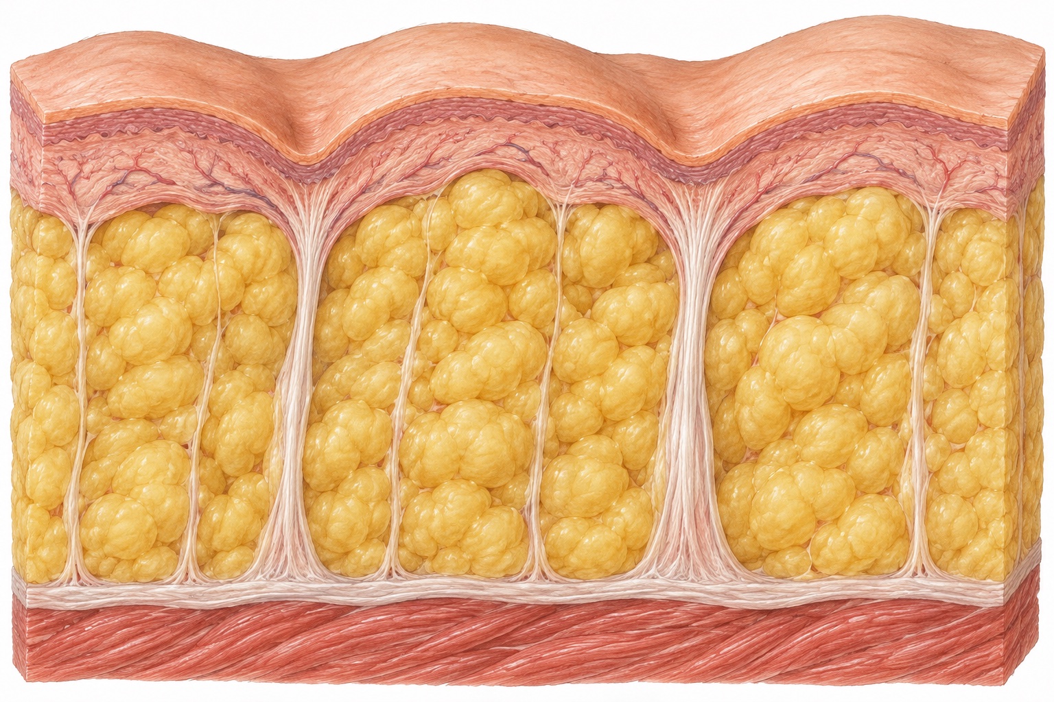
An MRI study of 30 women with cellulite on the buttocks compared dimpled areas with non-dimpled areas and found fibrous septa running perpendicular to the skin in 96.7% of dimpled sites, with the septa at dimpled sites much thicker than those at non-dimpled sites (Hexsel DM et al., 2009). Another MRI study similarly found that where cellulite was present, fat extended deeper into the dermis and a higher proportion of septa stood perpendicular to the skin (Querleux B et al., 2002). That is why cellulite commonly appears even in people with a lean body type and low body weight.
What's commonly called cellulite is a change in skin contour, but cellulitis is an acute infection in which bacteria invade the skin and subcutaneous tissue. If one leg suddenly becomes red and swollen and feels hot and painful to the touch, or if you have a fever throughout the body, this is not cosmetic cellulite. In that case, a bacterial infection or vascular problem should be checked first. So you should get immediate medical care, not cosmetic care.
Could Thick, Lumpy Thighs Be Lipedema?
Thighs that look lumpy or unusually thick can result not only from cellulite but also from lipedema or lymphedema, and the management approach differs because the underlying causes differ. Lymphedema is a condition in which lymphatic fluid fails to drain properly through the lymphatic vessels, causing the arms or legs to swell. Lipedema, by contrast, typically begins during periods of hormonal change in women, such as puberty, childbirth, or menopause, and is characterized by a symmetric increase in subcutaneous fat only in the arms and legs on both sides, while the hands, feet, and trunk stay the same (Torre YS et al., 2018).
When fat builds up due to lipedema, it feels like small nodules to the touch, and even light pressure can easily cause pain (Torre YS et al., 2018). Lipedema is frequently misdiagnosed as simple obesity or lymphedema. In addition, the increased leg fat generally does not go down easily with diet control and exercise alone.
Ultrasound measurement of skin and subcutaneous tissue thickness is one method used to tell different types of leg swelling apart. A review of 31 studies that examined ways to evaluate leg swelling by dividing it into general edema, lymphedema, and lipedema identified ultrasound as a useful test for differentiating lipedema, and presented it as a practical alternative particularly for obese patients or situations where MRI is difficult to perform (Markarian B et al., 2024). In fact, in a study that used ultrasound to examine the legs of 89 women, the 63 women with lipedema showed dermal and subcutaneous tissue thickness at the front of the thigh, front of the shin, and outer calf that differed clearly from the normal group, and the researchers proposed measuring thickness at these sites as an aid to ultrasound diagnosis (Amato ACM et al., 2021). Before treatment, THE WELLS first uses ultrasound to examine the thickness of the subcutaneous fascia in the thigh and how hardened it is.
Creams and Massage Devices Don't Reach the Septa
With self-care tools, the depth the stimulation reaches and the nature of the tissue it acts on are different, so they can't easily change the deep contours on the back of the thighs. Creams are applied to the epidermis, the outermost layer of skin, and home massage devices only press and rub the skin and the shallow fat layer just beneath it. The fibrous septa, by contrast, sit deep within the fat layer, holding the skin downward and binding it tightly to the fascia.
A review that synthesized prior research also noted that cellulite treatments targeting only the fat or the dermis showed effects that didn't last and produced inconsistent results, and concluded that the fibrous septa play a central role in cellulite formation (Bass LS et al., 2020). In fact, an analysis of 67 studies covering products and treatments found 19 rigorously designed randomized, placebo-controlled trials, and among these, the treatments with a possibility of meaningful effect were acoustic wave therapy in the shockwave category and some laser treatments (Luebberding S et al., 2015).
More recently, a study pooled data from 2,084 patients across 24 randomized controlled trials to compare the outcomes of treatments such as massage, topical treatments, shockwave therapy, and radiofrequency. This analysis also found differences depending on the treatment method (Lim SK et al., 2025). Shockwave therapy consistently lowered cellulite severity scores across four studies, and radiofrequency treatment reduced thigh circumference and subcutaneous tissue thickness (Lim SK et al., 2025).
We Check the Fascia With Ultrasound and Use Shockwave and Radiofrequency Together
Feeling the skin from the outside alone makes it hard to tell how hardened the subcutaneous tissue has become. So we first check body shape and body composition, then use ultrasound to assess the thickness and degree of hardening of the subcutaneous fascia in the thigh. The septa inside the subcutaneous fat layer and the condition of the fascia aren't visible to the eye, so we need to assess them with ultrasound to accurately set the depth of stimulation and the energy configuration.
After the ultrasound check, we apply two devices with different mechanisms in sequence. First, we use REFTONE extracorporeal shockwave therapy, a device that stimulates tissue through physical pressure waves rather than heat. For this, we use focused shockwaves that target deep focal points together with radial shockwaves that cover a wider area. REFTONE extracorporeal shockwave therapy reaches deep layers without damaging the skin surface, directly stimulating firmly hardened fibrous tissue and knotted areas. Next, we use WINBACK radiofrequency, a device that uses the TECAR method to generate biological heat deep within the body.
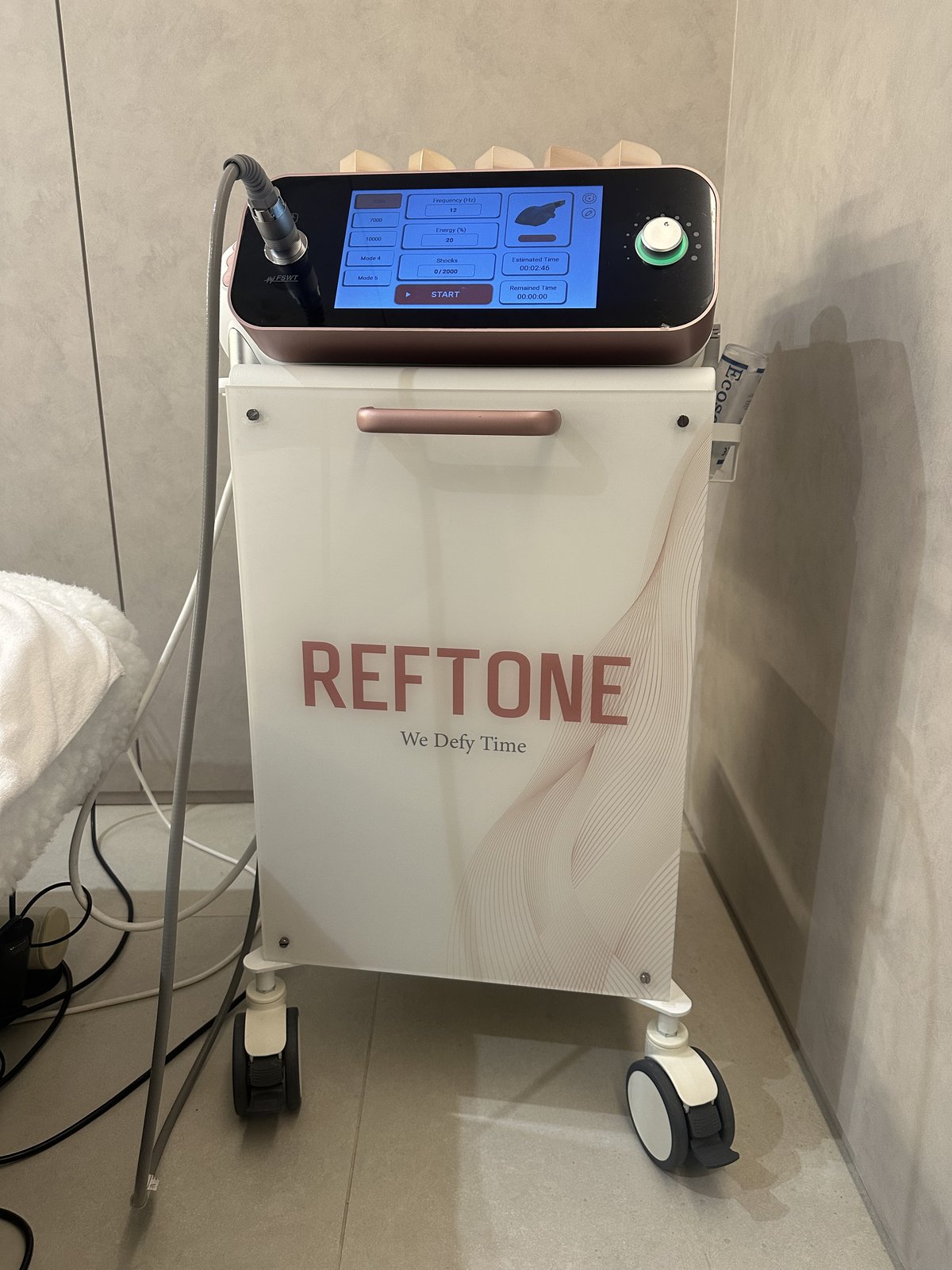
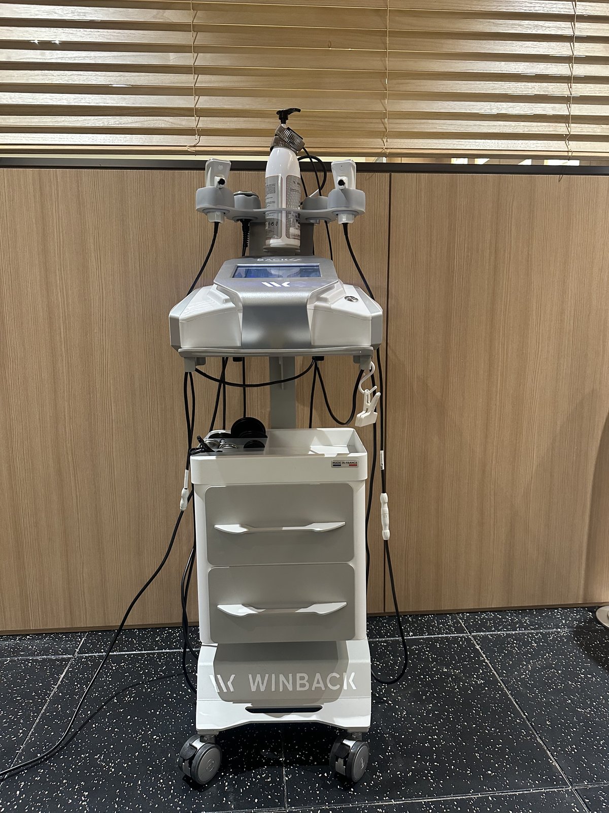
After stimulating the hardened layer with REFTONE extracorporeal shockwave therapy, we warm that deep layer with WINBACK radiofrequency. Stimulating the hard tissue first with physical pressure waves and then warming the deep layer causes blood vessels to dilate and relieves blocked circulation. Because the two devices do different jobs, we use them together.
In one randomized controlled trial, women with cellulite received focused extracorporeal shockwave therapy six times at 1- to 2-week intervals, combined with daily gluteal strengthening exercises. After 12 weeks, their severity scores dropped from 10.9 to 8.3, while a control group that did the same exercises with sham shockwave treatment showed no meaningful change (Knobloch K et al., 2013). Based on this evidence, when we carry out this combined care, our goal is to release the hardened layer that holds the skin on the back of the thigh downward and to improve circulation in the deep layers of the leg.
Will Shockwave and Radiofrequency Treatment Hurt?
During the procedure, shockwave therapy often feels like a firm, tapping sensation in the tissue, while radiofrequency tends to feel like warm heat spreading deep beneath the skin. Neither process is a surgical approach that involves cutting the skin or inserting needles. So you can return to your normal daily activities right after the procedure.
That said, areas where the subcutaneous fascia is especially stiff or where swelling is severe may feel more sore when the shockwave pulses reach them. We watch the response of the person receiving the treatment in real time and finely adjust the shockwave's impact pressure and the radiofrequency's heat intensity. After the shockwave and radiofrequency steps are finished, we follow with lymphatic circulation care to help the swelling pooled around the stimulated tissue and in the legs drain out smoothly.
The thickness of fat on the back of the thighs, the pulling force of the fibrous septa, and the usual degree of leg swelling differ from person to person. We set the combination and intensity of shockwave, radiofrequency, and circulation care, as well as the care schedule, one-on-one based on the initial ultrasound findings and the condition of each area. The total number of sessions varies depending on the area, the condition of the fascia, and lifestyle habits such as prolonged standing or sitting. So we periodically measure thigh circumference and changes in body shape and adjust the schedule.
Related Articles
References
- Luebberding S, Krueger N, Sadick NS (2015). Cellulite: an evidence-based review.. Am J Clin Dermatol. PMID: 25940753
- Hexsel DM, Abreu M, Rodrigues TC, Soirefmann M, do Prado DZ, Gamboa MM (2009). Side-by-side comparison of areas with and without cellulite depressions using magnetic resonance imaging.. Dermatol Surg. PMID: 19614939
- Querleux B, Cornillon C, Jolivet O, Bittoun J (2002). Anatomy and physiology of subcutaneous adipose tissue by in vivo magnetic resonance imaging and spectroscopy: relationships with sex and presence of cellulite.. Skin Res Technol. PMID: 12060477
- Bass LS, Kaminer MS (2020). Insights Into the Pathophysiology of Cellulite: A Review.. Dermatol Surg. PMID: 32976174
- Lim SK, Gultekin G, Suresan S, Jacob A, Zou Y, Liyanage DD (2025). Comparative Analysis of Cellulite Treatment Modalities: A Systematic Review.. Aesthetic Plast Surg. PMID: 39547984
- Knobloch K, Joest B, Krämer R, Vogt PM (2013). Cellulite and focused extracorporeal shockwave therapy for non-invasive body contouring: a randomized trial.. Dermatol Ther (Heidelb). PMID: 24297647
- Torre YS, Wadeea R, Rosas V, Herbst KL (2018). Lipedema: friend and foe.. Horm Mol Biol Clin Investig. PMID: 29522416
- Markarian B, Toro C, Moreira K, Polam S, Mathew N, Mayrovitz HN (2024). Assessment Modalities for Lower Extremity Edema, Lymphedema, and Lipedema: A Scoping Review.. Cureus. PMID: 38601427
- Amato ACM, Saucedo DZ, Santos KDS, Benitti DA (2021). Ultrasound criteria for lipedema diagnosis.. Phlebology. PMID: 33853452
Frequently Asked Questions
Q. Why does thigh cellulite appear even in a thin body type?
Even with low body fat, dimpling can appear depending on the structure of the fibrous septa that pull on the skin and the surrounding subcutaneous tissue. Rather than basing the management direction on body weight alone, it is necessary to check both the location of the skin depressions and the structure of the subcutaneous tissue.
Q. How are the number and interval of cellulite care sessions determined?
THE WELLS determines the number of sessions and the interval between them based on the condition of the subcutaneous tissue confirmed by ultrasound and on changes in tenderness and swelling after care. If soreness or swelling remains before the next session, we examine the area and then adjust the schedule and stimulation intensity.
Q. How should before-and-after photos of cellulite care be taken and compared?
Photos are taken under the same lighting and at the same distance, with the standing posture and the camera angle kept the same. Flexing the thigh or pressing on the skin can make the contours show differently, so the shooting conditions are kept constant and changes in the dimpled areas are compared.
Medical review: Jumin Kim · Medical Director · THE WELLS